Imaging of the Exposed Brain
Imaging of the Exposed Brain
As for NIR spectroscopy of brain, functional activation of cortical tissue can be monitored by reflectance monitoring and imaging and laser-Doppler spectroscopy and speckle imaging. All projects are based on our instrument developments and linked to the spectroscopic algorithms.
Neurovascular Coupling and Monitoring of Cortical Activation:
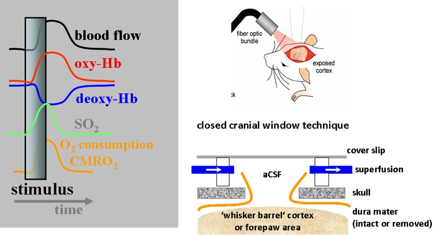
After neuronal activation by a stimulus the higher metabolic rate of oxygen (CMRO2) results in a vascular response with an increase in blood flow to match the oxygen consumption. Under normal conditions, this blood flow response over-compensates the oxygen extraction from the blood, with the effect of an increase in oxygenated haemoglobin and decrease in deoxygenated haemoglobin (neurovascular coupling). On the exposed cortex, the following parameters are accessible:
- Blood flow by laser- Doppler spectroscopy (1D) or Laser-Doppler imaging (2D) as well as speckle imaging (2D),
- Haemoglobin by reflectance monitoring (1D) or imaging (2D). In the lower part of the figure the experimental conditions of the exposed cortex with a cranial window is outlined.
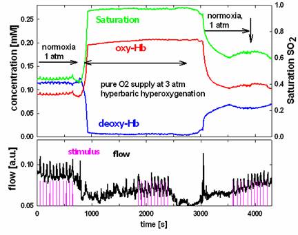
Example for the Monitoring of Cortical Activation - Activation during Hyperbaric Hyperoxygenation:
In the figure the recorded traces of the haemoglobin parameters and the blood flow is shown for three phases of 10 stimuli each (magenta), with the middle stimulus bout during hyperbaric hyperoxygenation (3 atm pure oxygen). The blood flow response (bottom part of the figure) is somewhat decreased. The haemoglobin response, however, is strongly suppressed during the hyperbaric period.
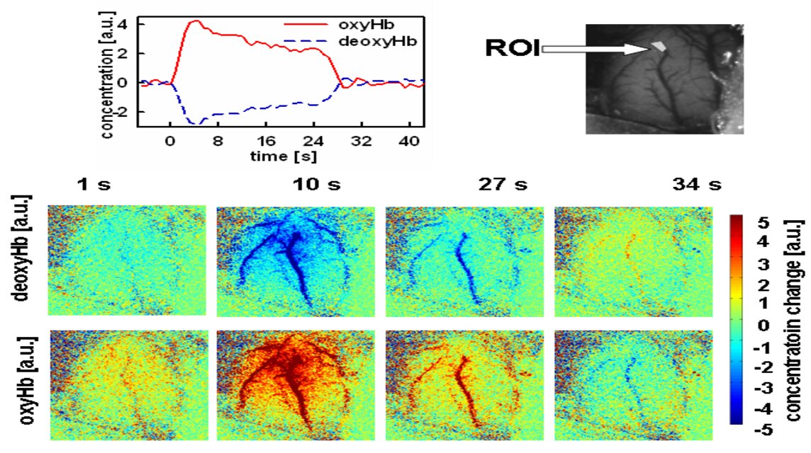
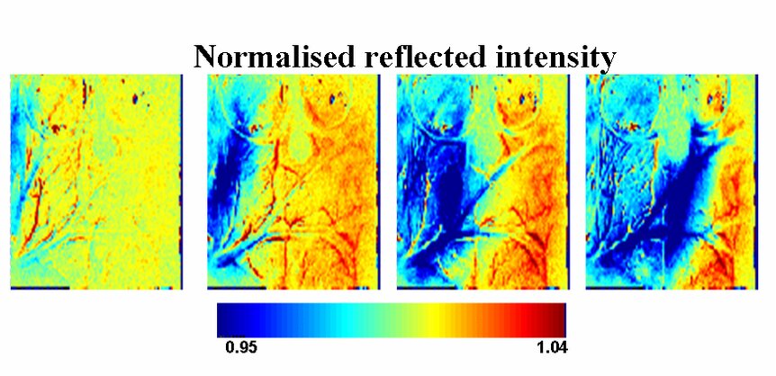
Imaging of Cortical Activation by combined Haemoglobin and Blood Flow Mapping:
Optical imaging based on a number of wavelengths is used for a mapping of cortical function in animal. The example shows both the time course of haemoglobin changes in a region of interest (ROI) and maps of calculated concentration changes for different times after cortical activation (time = 0 - 24 s).
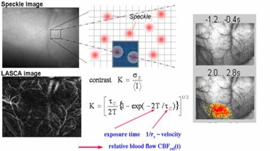
Speckle imaging is used to map the blood flow response in exposed cortical tissue.
Need for Instrumentation - Combined Haemoglobin and Blood Flow Imager:
A system combining both haemoglobin and blood flow imaging is under construction. The objective is to deliver a system with an online display of all parameters which is adapted for use by neuroscientists.
Optical Imaging for Stroke Research:
Both stroke and migraine is widely believed to be associated with a phenomenon called cortical spreading depression (CSD), which is a wave of neuronal activation and deactivation moving over the cortex. The figure shows a sequence of changes in haemoglobin on the cortex of a rat during CSD. Optical imaging and spectroscopy serves as a tool for stroke research.
Calculation of Cerebral Metabolic Rate of Oxygen CMRO2:
For cortical tissue the CMRO2 describes the effective metabolism of oxygen in the tissue and can be obtained from the spectroscopic data of haemoglobin and blood flow. With our instrumentation we study alterations of CMRO:
- by the intracranial pressure
- by pharmacological uncoupling of blood flow and metabolism, and
- hyperbaric hyperoxygenation.