Introduction to Tissue Optics
What is tissue Optics?
Tissue Optics describes the interaction of light, treated either as photons or waves, with biological tissue. In particular, the transport of light is studied in bulk tissues like brain, muscle or skin or teeth. The aim is the mathematical description of measurement parameters like intensity, time-of-flight, polarization or coherence.
What can be measured?
The objective is to measure absorbers (chromophores) and their concentrations scattering properties. blood flow, and tissue structure.
In biological tissue the absorption is dominated by a few chromophores, i.e.:
- water,
- haemoglobin and myoglobin (in muscle),
- lipids,
- aminoacids,
- melanin.
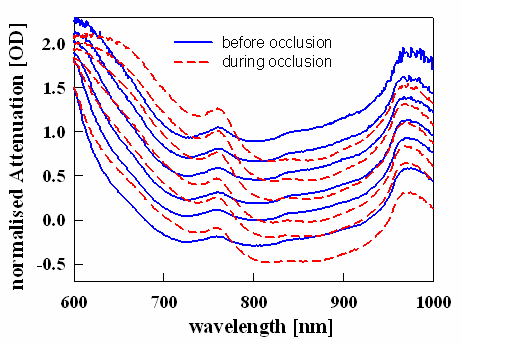
When looking at the absorption spectra over the Vis - NIR range (see figure) it is apparent that the absorption is low for wavelengths from 600 - 900 nm. Therefore, in this range the penetration into tissue is large (optical window).
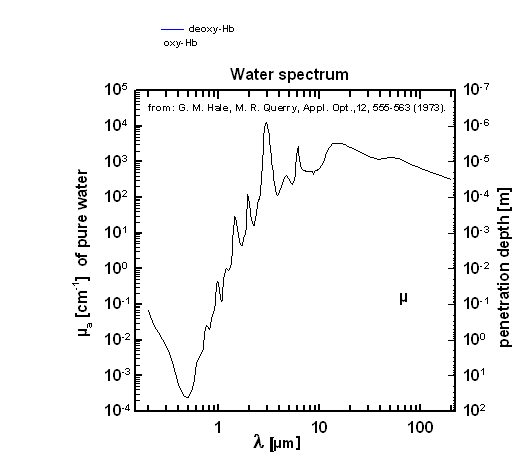
When looking at the water absorption spectrum over a larger wavelength range this optical window is even more apparent. The penetration depth, i.e. in inverse of the absorption coefficient, is smaller than 1 mm for all wavelength larger than about 1 µm.
What are the challenges of tissue optics?
- The tissue is inhomogeneous, both in its scattering properties and the distribution of chromophores.
- By and large, the scattering properties are unknown.
- For different tissues the background absorption from chromophores other than those mentioned above is unknown.
- The light intensity detected might be very low due to the light distribution in tissue.
How is the transport of light in tissue described?
The distribution of light in tissue is described by transport equations which result in what is called the diffusion regime for larger tissues. The key parameters for this description are
- the scattering coefficient µs which is the inverse between scattering events (unit: mm-1),
- the absorption coefficient µa which describes the likelihood of an absorption per path (unit: mm-1),
- the scattering angle, which is often described by its phase function or its mean cosine g,
- the transport scattering coefficient µs' = µs •(1-g),
- the refractive index n.
For simplest geometries (infinite or semi-infinite media, spheres) there are analytical equations which describe the intensity of the light as a function of distance from the light source, its time-of-flight etc. For other geometries or inhomogeneous media of a more complicated structure, numerical methods are used, predominantly Monte Carlo simulations.
What is the range of the optical tissue parameters?
- absorption coefficient µa: values strongly depend on the tissue and wavelength, typically in the range of < 0.01 mm-1 to 1 mm-1.
- the scattering coefficient µs: typically in the range of 5 – 20 mm-1, smoothly decreasing with wavelength.
- µs’ is typically in the range of 0.5 – 2 mm-1.
- n of tissue in about 1.4.
Which methods are used in tissue optics?
- Modified Labert-Beer method
- Time-domain-spectroscopy
- Intensity modulated spectroscopy
- Spatially-resolved spectroscopy
- Second-differential spectroscopy.
- Polarization of light is another measure for smaller source-detector separations.
- Laser-Doppler spectroscopy and laser speckle imaging are used for blood flow measurements.